The blood circulation of the apple snail is a typical example of the circulation in a monocardia: there is only one auricle that receives oxygen rich blood from the lung and the gills and deoxygenated blood from the kidney. So there is no separated blood circulation for oxygen rich and deoxygenated blood like in mammals and birds. It's a less efficient system, but it fulfils the needs of a snail very well.
The blood of apple snails (and snails in general) has two functions: transport of O2, CO2, hormones, nutrition and waste products and a structural function: a hydroskeleton.
The transport capacity of the blood for O2 and CO2 is enhanced by the chemical substance hemocyanine in the blood cells. Hemocyanine fulfils the same function as haemoglobin does in mammals (binding O2 and CO2 to ease transportation), but is colourless in contrary to the red colour of haemoglobin.
As the body of a snail does not contain a skeleton to support the extension movements, for example stretching out a tentacle, snails have to use another way: regulating the blood pressure in the body parts. In other words: inflating and deflating parts of the body in combination of muscle contraction to change shape. The regulation of the local blood is obtained by controlling the input and output of the bloodflow by contracting and relaxing small muscles that surround the veins.
Retracting movements are done by simple muscle contraction, without the need of fluid transportation.
| Movies (MPEG1): - Heartbeat seen through shell (Pomacea canaliculata) (97kb) |
| Snail heart in action: | |
 |  |
| Pomacea canaliculata. | |
The transport of the blood to and from the organs occurs through arteries (from heart to organs) and veins (from organs to heart). Snails don't have capillary veins and arterioles, which means their blood doesn't flow within tube-like structures (veins and arteries) during the whole circulation, but at the tissue level the blood circulates free between the cells and structures embedded in blood cavities (hemocoels) within the body (=open circulation).
The circulation and filtration of the blood:
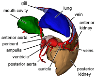
The heart of apple snails is well developed and consists of two chambers: the auricle and the ventricle.
The auricle is which receives the blood influx from the lung and the kidney veins is much smaller then the ventricle. Inside the auricle, there are many small muscle fibres connecting the opposite wall trough the lumen. The walls have a spongy surface at the inside and a basal layer at the outside. The blood is able to reach the basal membrane through the spongy surface. Contraction is achieved by contracting these muscle fibres.
The ventricle is much larger and has thick, muscular walls with many spaces between the wall muscles, allowing the blood to be trapped in these semi-vescicles and filtered through the basal membrane. In contrast with the auricular contraction, the ventricular contraction is based on contraction of the wall muscles. The mean ventricular pulse pressure of the African apple snail Lanistes carinatus is reported to be around 7.8 cm of water.
The aortic ampulla functions as a compensation sac and compensates the elevated blood pressure in the aorta during the contraction of the ventricle. Besides its function to regulate the blood pressure, the ampulla also has a function in the immune system as the walls of the ampulla consists of vacuolated tissue with many phagocytes in it. These phagocytes possibly eliminate micro-organisms from the bloodflow. The wall of the ampulla is relatively impermeable, excluding the ampulla for blood filtration.
Both the heart and the ampulla are embedded in the pericard, which is connected with the posterior kidney through a renopericadial canal. The walls of the pericard cover the heart and the ampulla and in the pericard cavity the walls are covered with microvilli, small intercellular channels and ridges. Near the renopericardial canal there are some mucous cells secreting mucus, presumably to bind small particles to be transported to the kidney.
The fluid excreted in the pericard cavity can be considered to be primary urine and this fluid is transported to the kidneys for further filtration and resorption of usable compounds (sodium and chlorine).
The kidney or nephridium consists of two parts: the posterior chamber which excretes uric acid and purines and the anterior chamber which has osmoregulatory function.
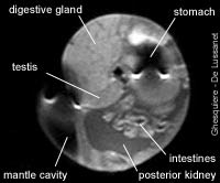 MRI image of a male apple snail (Pomacea diffusa). |
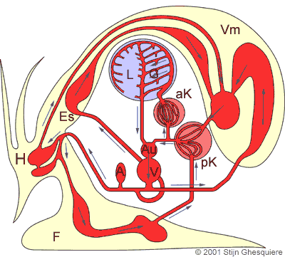
The heart action:
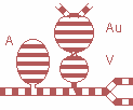 Au = Auricle V = Ventricle |
The posterior kidney chamber receives the primary urine from the pericard cavity. The folds on the posterior chamber wall have a dense vascular network and are covered with excretory, ciliated and mucous cells. The excretory cells excrete uric acids and other purines from the blood into the lumen of the chamber in which the primary urine flows.
The anterior kidney chamber differs from the posterior chamber in that the lumen is occluded with large lamina that covers the walls. These lamina remarkably increase the surface area that comes in contact with the urine. The epithelium on these lamina is almost entirely consisting of resorptive cells that presumable resorb ions from the urine into the blood.
The renal aperture (urine opening) is situated in the upper region of the right mantle cavity. The urine produced by the kidneys is expelled here.
The aorta with it's white calcareous granula in it's wall consists two parts: the anterior and the posterior part.
The anterior aorta connects the heart with the head (cephalic hemocoel) and the foot (foot hemocoel), while the posterior aorta divides close to the heart with one artery distributing the blood to the digestive system and the second serves several other organs (testis, ovaria, intestines etc.).
After circulating through the tissues and hemocoels of the snail, the blood is collected in large veins and brought to the posterior kidney.
A portion of the blood that enters the posterior kidney directly flows back to the heart, while the remaining blood enters the vascular system of the kidneys (posterior and anterior part).
After passing the anterior kidney, the blood flows through the mantle vein, from where many small veins bring the blood to the gills and the lung, where O2 uptake and CO2 is exchange takes place.
The lung-gill vein collects the oxygen rich blood from the lung and the gills and brings it back to the heart.
No comments:
Post a Comment