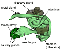
The digestive system of apple snails is adapted to feed on aquatic plants.
Roughly it can be in five regions: the intake region (mouth and buccal cavity with radula and jaws), the pre-digestion region (oesophagus with salivary glands, lateral pouchs and crop), digestion region (three chambered stomach with the associated digestive gland), uptake region (intestine) and the excretion region (rectum and anus).
Roughly it can be in five regions: the intake region (mouth and buccal cavity with radula and jaws), the pre-digestion region (oesophagus with salivary glands, lateral pouchs and crop), digestion region (three chambered stomach with the associated digestive gland), uptake region (intestine) and the excretion region (rectum and anus).
| Interactive 3d-model | Scheme of the digestive tract |
 Interactive 3D-model of the digestive tract of a Pomacea canaliculata snail. Click on picture to interact Note: can take some time to load. | 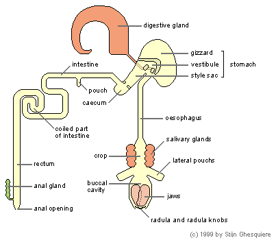 |
The mouth of the apple snail is a vertical slit opening, located between the labial tentacles and leading to the buccal cavity.
Food is located with the labial tentacles and when needed gathered from the water surface with foot.
Food is located with the labial tentacles and when needed gathered from the water surface with foot.
 The radula of an eating snail in detail (Pomacea canaliculata). (click on image to enlarge) |
|

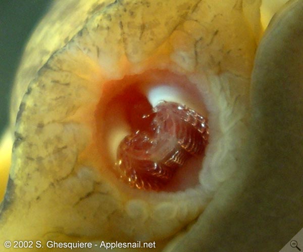
The buccal cavity, a muscular cavity with a set of calcareous jaws, 2 radula knobs and the radula (rasp tongue), is situated behind the mouth opening.
The radula lies on top of the radula knobs (odontophores) and is covered with several rows (26 to 53), each consisting of 7 renewable chitinous teeth. When the radula knobs are moved from each other, the radula is bend and stretched, this spreading the teeth on it, which provides the grasp function.
After the food has pulled into the buccal cavity, the strong, calcareous jaws cut off the piece near the mouth opening.
More details about the mouth movements here.
| Movies (MPEG1): - Mouth in eating apple snail (Asolene megastoma) (202kb) |
Due to the myglobin rich muscles, the buccal mass (the complete organ with the cavity within) has a reddish colour. Myoglobin is a respiratory pigment that helps to provide sufficient oxigen for the muscle metabolism during activity. The buccal mass itself lies in the anterior part of the cephalic hemocoel (a large vascular sinus or cavity).
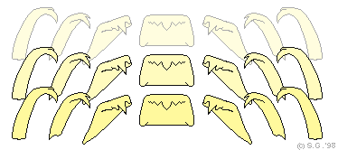 Rows of the teeth (7) on the radula (Pomacea canaliculata). | 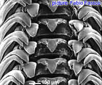 Microscopic picture of the radula from Felipponea neritiformis. |
At the back of the buccal cavity there are two lateral pouches and the ducts of the salivary glands are located here as well. The bright orange salivary glands lie against the expanded first part of the oesophagus (the crop) and produce an acid mucopolysaccharide.
The walls of the crop and the oesophagus are covered with longitudinal folds and contain cells that excrete a neutral mucopolysaccharide and proteins in addition. The oesophagus connects the buccal cavity with the stomach after making a turn of 180° (following the coiled body of the snail). The food is transported through the oesophagus with peristaltic movement.
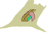
The stomach of the apple snail is a pink; "U" shaped structure on the left side of the body whorl. The wall of the stomach consists of several muscular layers, often covered with calcareous deposits of the overlaying tissues or by the digestive gland.
The stomach itself consists of three compartments: the muscular gizzard (posterior chamber), the vestibule (anterior chamber) and the style sac.
The walls of the crop and the oesophagus are covered with longitudinal folds and contain cells that excrete a neutral mucopolysaccharide and proteins in addition. The oesophagus connects the buccal cavity with the stomach after making a turn of 180° (following the coiled body of the snail). The food is transported through the oesophagus with peristaltic movement.
The stomach of the apple snail is a pink; "U" shaped structure on the left side of the body whorl. The wall of the stomach consists of several muscular layers, often covered with calcareous deposits of the overlaying tissues or by the digestive gland.
The stomach itself consists of three compartments: the muscular gizzard (posterior chamber), the vestibule (anterior chamber) and the style sac.
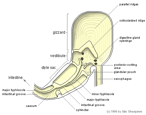 |
The food is mixed with digestive enzymes and slime with the muscular movements in the gizzard and which connects with vestibule.
The gizzard and the vestibule are separated by a thick ridge that reaches to the roof of the stomach, closing the gizzard off. When the muscles of the gizzard contract, the gizzard and the vestibule communicate. The surface of the gizzard had many parallel folds (ridges) and the walls consist of several muscular layers.
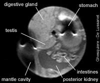
The ducts of the digestive gland or midgut gland, which produces digestive enzymes, opens in the ciliated vestibule. The midgut gland lies on top of the stomach and in line with the testis/ovaria in the upper whorls of the shell. The function of the midgut gland might be comparable with the human liver (biochemical conversions of nutrients and waste) combined with the pancreas (production of digestive enzymes), and is also called the hepatopancreas. One can recognize the midgut gland easily due to it's dark brown colour.
The gizzard and the vestibule are separated by a thick ridge that reaches to the roof of the stomach, closing the gizzard off. When the muscles of the gizzard contract, the gizzard and the vestibule communicate. The surface of the gizzard had many parallel folds (ridges) and the walls consist of several muscular layers.
The ducts of the digestive gland or midgut gland, which produces digestive enzymes, opens in the ciliated vestibule. The midgut gland lies on top of the stomach and in line with the testis/ovaria in the upper whorls of the shell. The function of the midgut gland might be comparable with the human liver (biochemical conversions of nutrients and waste) combined with the pancreas (production of digestive enzymes), and is also called the hepatopancreas. One can recognize the midgut gland easily due to it's dark brown colour.
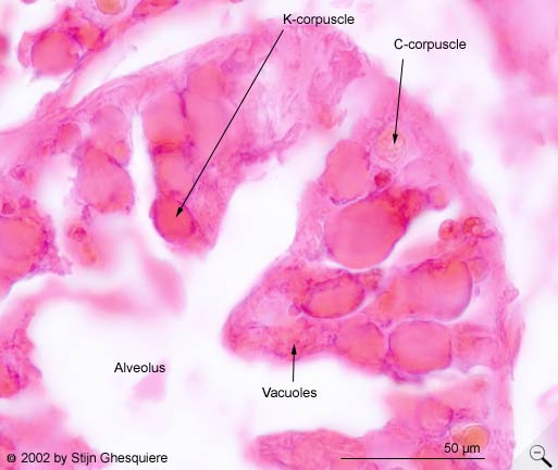
 C and K-corpuscles from the midgut gland of Pomaceacanaliculata. Click on image to enlarge. |
Inside the digestive or midgut gland of snails from the genera Pomacea, Marisa and Asolene and most likely in all ampullarids, a large quantity of a greenish-black substance is present. When studied under a microscope, two types of pigmented elements can be observed: cellular round corpuscles (C corpuscles) with a brownish/greenish colour and dark brown 'club' shaped elements (K or kystic corpuscles). The C corpuscles measure about 11 - 13 µm in diameter and the K corpuscles are about 35µ in lenth and 13µm in width. Both corpuscles are also present in the intestines and the feces. They even show up in debris of the snail's habitat.
Both corpuscles types are not only found in the ducts and the digestive tract, they also reside inside the glandular cells. The C elements reside in columnar cells, which are considered to be digestive cells and the K elements are associated with the supposedly excretory pyramidal cells. In total these corpuscles make up a considerable amount of the glandular mass (around 11-13% of the total gland mass).
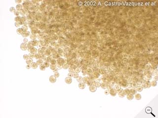
The corpuscles are excreted from the gland cells in the gland ducts and are transported to the stomach, where they mix with the food. The corpuscles are present in the feces as well, where they are intermingled with the food remnants. Besides leaving the snail's body with the normal feces, the corpuscles are also excreted in dense droppings, consisting of nearly 100% corpuscles (see this picture of a Pomacea insularum dropping and a similar one from a Pomacea canaliculata dropping). These droppings contain mainly the C corpuscles and much less K corpuscles, while the normal feces mainly contain K-corpuscles.
Recent research [VEGA 2001 and CASTRO-VAZQUEZ, 2002] revealed that the C and K corpuscles might be a symbiotic (prokaryotic) organism, with the K corpuscles being the kystic form of the vegetative C elements. The fact that these elements contain DNA and have a double external membrane, but the lack of a nucleus supports the view of a prokaryotic origin of these corpuscles.
The putative symbiont appears to be well integrated in the snail as even sterile raised hatchlings contain these corpuscles, excluding an external alimentairy origin. Also the observed intracellular presence and development suggest a close relationship. The C corpuscles are for example derived from spheric bodies (vacuoles) that develop and divide inside the digestive columnar cells. These unsheated vacuoles are excreted into the alveoli and develop into mature C-corpuscles. During their travel through the intestines the C-corpuscles become sheated and transform into K-corpuscles.
A symbiont living inside these snails implies that the snail and the symbiont (the prokaryote) have established a mutual beneficial relationship somewhere in the course of evolution. For the putative symbiont, the benefits are clear: a protective and food enriched environment. For the snail the positive effects aren't that clear yet. For example, the snail looses valuable nutritional proteins as these are partly used by the symbiont, which becomes excreted with the feces. Possibly a biochemical advantage is given to the snail in this relationship. Further research will certainly reveal more of this interesting symbiotic relationship
Both corpuscles types are not only found in the ducts and the digestive tract, they also reside inside the glandular cells. The C elements reside in columnar cells, which are considered to be digestive cells and the K elements are associated with the supposedly excretory pyramidal cells. In total these corpuscles make up a considerable amount of the glandular mass (around 11-13% of the total gland mass).
The corpuscles are excreted from the gland cells in the gland ducts and are transported to the stomach, where they mix with the food. The corpuscles are present in the feces as well, where they are intermingled with the food remnants. Besides leaving the snail's body with the normal feces, the corpuscles are also excreted in dense droppings, consisting of nearly 100% corpuscles (see this picture of a Pomacea insularum dropping and a similar one from a Pomacea canaliculata dropping). These droppings contain mainly the C corpuscles and much less K corpuscles, while the normal feces mainly contain K-corpuscles.
Recent research [VEGA 2001 and CASTRO-VAZQUEZ, 2002] revealed that the C and K corpuscles might be a symbiotic (prokaryotic) organism, with the K corpuscles being the kystic form of the vegetative C elements. The fact that these elements contain DNA and have a double external membrane, but the lack of a nucleus supports the view of a prokaryotic origin of these corpuscles.
The putative symbiont appears to be well integrated in the snail as even sterile raised hatchlings contain these corpuscles, excluding an external alimentairy origin. Also the observed intracellular presence and development suggest a close relationship. The C corpuscles are for example derived from spheric bodies (vacuoles) that develop and divide inside the digestive columnar cells. These unsheated vacuoles are excreted into the alveoli and develop into mature C-corpuscles. During their travel through the intestines the C-corpuscles become sheated and transform into K-corpuscles.
A symbiont living inside these snails implies that the snail and the symbiont (the prokaryote) have established a mutual beneficial relationship somewhere in the course of evolution. For the putative symbiont, the benefits are clear: a protective and food enriched environment. For the snail the positive effects aren't that clear yet. For example, the snail looses valuable nutritional proteins as these are partly used by the symbiont, which becomes excreted with the feces. Possibly a biochemical advantage is given to the snail in this relationship. Further research will certainly reveal more of this interesting symbiotic relationship
In the vestibule, in front of the style sac is a sorting area at which the large food parts are filtered out.
The entrance of the style sac is marked with a strong muscular sphincter, connected with the minor and the major typhlosole and between them a deep, longitudinal intestinal groove. On the typhlosoles is a strong ciliary current towards the intestine, while the walls of the style sac have a transverse rotating ciliary current (giving a spiral movement to the food).
 MRI image of a male apple snail (Pomacea diffusa). |  The dark line you can see through the shell is the rectum, leading to the anus (Pomacea diffusa). |
The digestion of the food is partly done by adding enzymes by the snail itself and partly by the micro-organisms in the intestine. The uptake of the food compounds is carried out trough the walls of the intestine and transported to the blood stream. The posterior aorta provides the blood supply of the intestine.
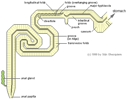 |
The ciliated major typhlosole, which starts in vestibule of the stomach with the intestinal groove parallel to it, continues throughout the caecum and the first part of the intestine. The walls of the first part of the intestine are covered with ciliated folds that transport the food. The intestinal groove in this part of the intestine doesn't have those fold or ciliation. At the end of the first part, the intestinal groove expands to a pouch and the typhlosole bends to the pouch, while its tip points to the shallow groove of the second part of the intestine.
The shallow, unciliated groove in the second part of the intestine is partly covered with ciliated folds. The epithelium lining the groove contains glands that produce an acid mucopolysaccharide, this in contrast with the neutral mucopolysaccharide produced elsewhere on the walls.
At the junction with the third part of the intestine (rectum) the groove is deflected in a small diverticle. The typhlosole (ridge) lining the groove becomes greatly enlarged and the groove becomes narrow and is located on top of the ridge. The walls of the third region of the intestine have unciliated transverse folds. Close to the anal opening intestine receives the duct of the anal gland, which arises at the base of the anal papilla.
The anal papilla is situated at the roof of the right mantle skirt.
| The mouth movements: When the snail eats, the buccal cavity if brought toward the mouth opening and then a complex mechanism of grasping and cutting starts. In the picture below you see a simplified version of the mouth movements in the apple snail. It might help you to get an idea of how it works. Note that the radula in the real apple snail is more or less covered by the radula knobs that bend and stretch the radula to get the radula teeth moving towards and away from each other in order to grasp the food. These knobs can be easily confused with the jaws. The jaws are more difficult to see, they only come out of the mouth a very little bit after the radula has pulled back in and the mouth is already closing.
At first, the mouth opens, while the jaws are lifted from the radula. The radula is then brought forward, while the teeth on the radula are spread. The radula moves like a caterpillar track over the underlying radula knobs. At the point that the radula is at its most forward position, the radula teeth bend toward each other and grasp the food. The radula is pulled back in, while the jaws are brought forward to cut off a piece of food, similar to the mechanism of a scissors. After the food piece has been cut off, it's brought to the oesophagus. | |
No comments:
Post a Comment